


KI News
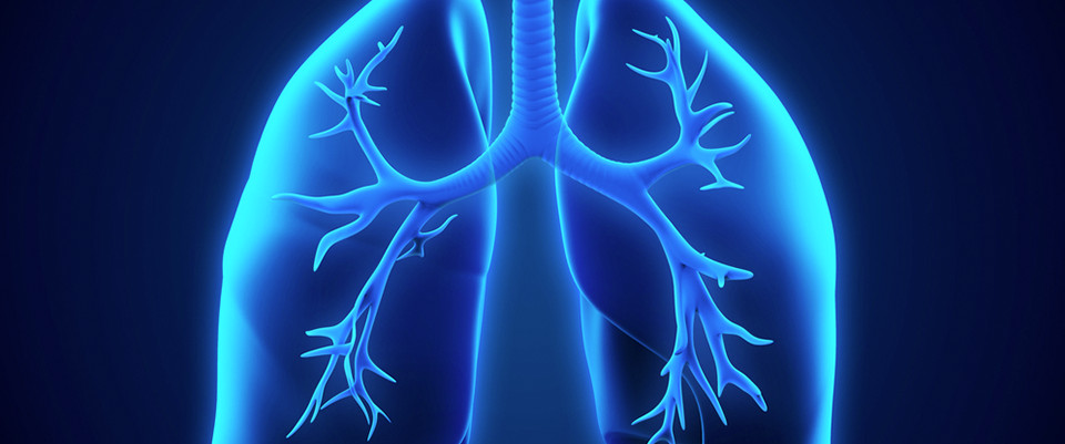
Oxygen therapy for patients suffering from a heart attack does not prevent heart failure
Oxygen therapy does not prevent the development of heart failure. Neither does it reduce the long-term risk of dying for patients with suspected heart attack. This has been proven for the first time by researchers at Karolinska Institutet as a result of a major Swedish study. The study is to be presented at the European Society of Cardiology’s (ESC) cardiology congress in Munich and published at the same time in the journal Circulation. The researchers expect their results to have a global impact on recommended healthcare for treating heart attacks.
Oxygen has been used to treat patients suffering a heart attack for more than a century, despite the fact that such treatment has not had any scientifically proven effect on patients who have normal oxygen levels in their blood. Since the turn of the millennium, researchers worldwide have started to question whether oxygen therapy for heart attacks is ineffective – or may even be harmful.
“Our new study has filled a central gap in knowledge regarding how to treat patients suffering a heart attack. One year ago, we were able to confirm that oxygen therapy does not appear to reduce the risk of dying up to one year after the heart attack. We can now substantiate these findings for a long-term perspective and show that oxygen therapy does not reduce the development of heart failure, the most worrying complication of heart attacks. On this basis, the routine use of oxygen can now be eliminated, and healthcare personnel can concentrate on more efficient measures and rapid transport to hospital,” confirms Robin Hofmann, senior consultant cardiologist and researcher at the Department of clinical science and education, Södersjukhuset, at Karolinska Institutet.
The DETO2X-AMI study was conducted at 35 Swedish hospitals, involving random treatment with or without oxygen of 6,629 patients with suspected heart attack. The result shows that oxygen therapy in a moderate dose is not harmful but does not increase the survival rates or reduce complications, such as the development of heart failure or new heart attacks.
The research project was financed by the Swedish Heart-Lung Foundation and the Swedish Research Council.
Publication
”Long-term effects of oxygen therapy on death or hospitalization for heart failure in patients with suspected acute myocardial infarction”
Tomas Jernberg, Robin Hofmann, et al for the DETO2X-SWEDEHEART investigators.
Circulation, 26 August, 2018, doi: 10.1161/CIRCULATIONAHA.118.036220
Treatment for severe heartburn prevents cancer
Medical or surgical treatment of severe heartburn prevents cancer of the oesophagus, a study from Karolinska Institutet with almost one million Nordic patients reveals. The results will be published in the scientific journal JAMA Oncology.
Pathological heartburn and acid reflux affects 10-20 per cent of the adult population. Long and severe reflux is the strongest risk factor for cancer of the oesophagus (type adenocarcinoma), an aggressive cancer that is difficult to treat.
Reflux is usually treated with medicine to make the stomach contents less acidic, which usually eliminates or reduces symptoms. One alternative is to have an operation (anti-reflux surgery) which prevents the stomach contents from coming up into the oesophagus. Previous studies have not conclusively demonstrated that these treatments prevent oesophageal cancer, but the studies have not been sufficiently large or had enough follow-up time to ensure that conclusions can be drawn on any long-term cancer-preventive effects.
940,000 patients with reflux included in the study
In the present study, researchers used health data records from 1964 to 2014 from the five Nordic countries. Of the more than 940,000 patients with reflux in the study, about 895,000 received medical treatment and of those nearly 2,370 patients (0.3 per cent) developed cancer of the oesophagus during the follow-up period. The risk of cancer of the oesophagus decreased over time following treatment and was similar to that of the corresponding population after 15 years or more in those who received medication.
Of the more than 48,400 patients who had anti-reflux surgery, 177 (0.4 per cent) developed cancer of the oesophagus during the follow-up period. The risk of oesophageal cancer clearly fell also in this group and was at the same level as in the corresponding population 15 years or more after the operation.
When the patients with reflux who had an operation were compared with those with reflux who received medication, the patients who had been operated on had a slightly higher risk of oesophageal cancer during the entire follow-up period, but the risk did not increase over time. This is probably caused by the fact that the operated patients had more serious reflux from the beginning.
“The results show that effective medical or surgical treatment of reflux prevents cancer of the oesophagus. But because the individual’s risk of developing oesophageal cancer is low, even in those with reflux disease, the results do not justify treating reflux solely as a cancer-preventive measure. The symptoms and complications of reflux disease should continue to govern treatment,” says John Maret-Ouda, physician and scientist at the Department of Molecular Medicine and Surgery at Karolinska Institutet and the first author of the study.
However, he points out that for the small percentage of people with severe reflux in combination with other risk factors for oesophageal cancer, such as obesity, male gender and mature age, effective and continuous medical treatment or an operation to treat reflux is recommended.
Statistically significant results
“Previous research results have shown poor cancer-preventive effects from anti-reflux surgery. The difference now is that for the first time we can show statistically significant results because we have a sufficiently large study with a long follow-up period of over 15 years following the operation,” says Jesper Lagergren, consultant surgeon and professor at the Department of Molecular Medicine and Surgery, Karolinska Institutet, who led the study.
The research is funded by the Nordic Cancer Union, the Swedish Research Council and the Swedish Cancer Society.
Publication
”The risk of esophageal adenocarcinoma following antireflux surgery in the five Nordic countries”
John Maret-Ouda, Karl Wahlin, Miia Artama, Nele Brusselaers, Martti Färkkilä, Elsebeth Lynge, Fredrik Mattsson, Eero Pukkala, Pål Romundstad, Laufey Tryggvadóttir, My von Euler-Chelpin and Jesper Lagergren.
JAMA Oncology, online 23 August, 2018, doi:
Dramatic development of immune system after birth
As soon as a baby is born, its immune system starts to change dramatically in response to the bacteria, viruses and so forth in its new environment, a phenomenon that is common to all babies, researchers from Karolinska Institutet in Sweden write in a paper published in Cell. The study was made possible using new techniques of immune cell analysis.
Examining how the neonatal immune system changes has been difficult since the analyses are made from samples taken from the umbilical cord directly after delivery. Researchers have now exploited a new technique of immune cell analysis to monitor how babies develop for the first few weeks of life outside the womb.
“This is the first time we’ve pinned down how the human immune system adapts itself to birth and the new environment,” says Petter Brodin, doctor and researcher at the Science for Life Laboratory (SciLifeLab) and the Department of Women’s and Children’s Health, Karolinska Institutet. “We saw drastic changes in the babies’ immune system between each sampling, which shows that it is highly dynamic early in life.”
The study compared blood samples from 100 babies, both premature and full-term, taken during the first, fourth and twelfth week. The comparison was achieved using an advanced technique of immune cell analysis: mass cytometry in combination with extensive plasma protein analyses.
Only small amounts of blood, just a few drops from each baby, are needed to analyse all the white blood cells and hundreds of proteins circulating in the blood. The researchers were also able to show that the babies who had abnormal development of the gut flora during the first weeks also demonstrated a disorder of the immune system.
“Our results are important for better understanding the infection-sensitivity of newborn babies and the risks of premature birth,” says Dr Brodin. “If we can monitor the development of the immune system and steer it in different directions, we make it possible to prevent autoimmune diseases and allergies, which are partly related to the development of the immune system, and to even develop better vaccines, tailored to the neonatal immune system.”
The adaptation of the immune system is thought to be triggered by the microbes, bacteria, fungi and so forth that the baby encounters outside the womb. The process begins primarily in the lungs, gut, skin and mucosa, which is to say the body’s points of contact with the outside world.
“What surprised us was how similar the changes were amongst babies,” says Dr Brodin. “It seems as if all babies follow one and the same pattern, with their immune systems responding with exactly the same sequence of dramatic changes. It’s almost like a well-choreographed dance, a practised routine.”
The researchers will now be broadening out the study to encompass more babies, all of whom will be monitored into childhood. This will enable the team to see which of them develop diabetes, allergies, asthma and inflammatory bowel disease.
“Many of these diseases can definitely be traced back to how a baby is born and how its immune system adapts to the external environment,” explains Dr Brodin. “What we’re bringing to the table is the specific changes in the immune system that underlie this. It’s a piece of the puzzle that was formerly missing.”
The study was made possible through the close collaboration of Karolinska University Hospital.
“For ethical, practical and logical reasons it’s difficult to put together a study like this,” says Dr Brodin. “The key to our success is that those of us leading the study also work as doctors and when we manage to combine patient-end work with the most advanced techniques, we make wonderful discoveries.”
The researchers who conducted the study are active at the Science for Life Laboratory, Karolinska Institutet, and Karolinska University Hospital.
The study was financed with grants from the European Research Council (ERC), the Swedish Research Council, Karolinska Institutet and the Swedish Society for Medical Research (SSMF).
Publication:
“Stereotypic immune system development in newborn children”, Axel Olin, Ewa Henckel, Yang Chen, Tadepally Lakshmikanth, Christian Pou, Jaromir Mikes, Anna Gustafsson, Anna Karin Bernhardsson, Cheng Zhang, Kajsa Bohlin, Petter Brodin. Cell, online 23 August 2018, doi: 10.1016/j.cell.2018.06.045.
Cleverly located surface proteins make some pneumococcal strains especially dangerous
Successful pathogenic strains of pneumococci have two proteins that, owing to their location on the surface of the bacteria, enhance their survival and ability to cause disease, a study from Karolinska Institutet in Sweden published in Nature Communications, reports.
Pneumococcal infections are one of the most common causes of disease and death in the world. One reason for the pathogenic potential of these bacteria is that they produce a sugar casing. This capsule prevents the important immune component C3b from attaching to and attacking the bacteria.
Researchers at Karolinska Institutet and the Royal Institute of Technology in Sweden have now studied in detail how pneumococci interact with the part of the immune system called the “complement system”, which includes C3b. The complement system often works as the first line of defence against foreign substances and cells, triggering a number of immune reactions in the body.
The researchers show that the capsule is weak at the bacteria’s point of division, which therefore presents an opening for C3b. By using super-resolution microscopy (STED) they found that C3b accumulates under the capsule primarily at the division sites. This accumulation can continue and cover the entire bacteria unless the pneumococcus can find a strategy to prevent it happening.
The study also shows that a common surface protein on pneumococci called PspC1 is located right at the division site, where it recruits another protein called Factor H, which negatively regulates the complement system by, amongst other mechanisms, inactivating C3b. Some especially successful and pathogenic pneumococcal strains also express a closely related protein, PspC2, which is mainly localised at the bacterial poles. This separate location on the surface of the bacteria affects the two surface proteins’ functions. Unlike PspC1, which binds Factor H, PspC2 affects the bacteria’s ability to adhere to epithelial cells, which can be found in the respiratory tract, in mucus membranes and elsewhere.
“Our study shows that the precise localisation of bacterial surface proteins in relation to the capsule layer affects the role they will have in the disease development,” says Birgitta Henriques-Normark, professor at the Department of Microbiology, Tumour and Cell Biology, Karolinska Institutet. “This is an important piece of the puzzle to understand how pneumococci avoid the immune system and cause everything from otitis and sinusitis to severe pneumonia and septicaemia.”
The study was financed by the Swedish Research Council, Stockholm County Council (ALF funding), the Swedish Foundation for Strategic Research and the Knut and Alice Wallenberg Foundation.
Publication
“Factor H binding proteins protect division septa on encapsulated Streptococcus pneumoniae against complement C3b deposition and amplification”. Anuj Pathak, Jan Bergstrand, Vicky Sender, Laura Spelmink, Marie-Stephanie Aschtgen, Sandra Muschiol, Jerker Widengren & Birgitta Henriques-Normark. Nature Communications, online 23 August 2018, doi: 10.1038/s41467-018-05494-w.
New scientific model reveals interaction between blood vessels and neurons in the brain
Micro-flow models of the blood-brain barrier and the brain reveal for the first time complex metabolic interactions between the brain’s blood vessels and nerve cells. The findings are published by an international team of researchers, including from Karolinska Institutet and the Royal Institute of Technology, in the journal Nature Biotechnology.
Interaction and regulation between blood vessels and nerve cells in the brain are essential to its function, but how this interaction takes place has been difficult to pin down. Classic cell-culture models of nerve cells and blood vessel cells are too simplified to show interactions, and studies on brain tissue and animals are too complicated.
In order to understand how blood vessels and surrounding brain tissue interact, the team built up a so-called “organ-on-chip”-model, which is a micro-flow model with living cells.
Using the model, the researchers were able to analyse all the smaller molecules issued by the cells and identify a previously undescribed link for how blood vessel cells metabolise glucose and how these metabolites influence the production of neurotransmitters by the neurons.
The team is now working on using these systems to obtain new insights on the brain and to build models of brain diseases.
The study was financed by the Wyss Institute for Biologically Inspired Engineering at Harvard University, Defense Advanced Research Projects Agency (DARPA), the Sweden-America Foundation, the Carl Trygger Foundation and the Erik and Edith Fernström Foundation.
Publication
“A linked organ-on-chip model of the human neurovascular unit reveals the metabolic coupling of endothelial and neuronal cells”
Ben M Maoz, Anna Herland, Edward A FitzGerald, Thomas Grevesse, Charles Vidoudez, Alan R Pacheco, Sean P Sheehy, Tae-Eun Park, Stephanie Dauth, Robert Mannix, Nikita Budnik, Kevin Shores, Alexander Cho, Janna C Nawroth, Daniel Segrè, Bogdan Budnik, Donald E Ingber and Kevin Kit Parker
Nature Biotechnology, online 20 August 2018, doi: 10.1038/nbt.4226
Karolinska Institutet keeps a leading position
The recently published Shanghai ranking (ARWU – Academic Ranking of World Universities) shows that Karolinska Institutet keeps a leading position as one of the world's strongest universities and the highest-ranked university in Sweden.
KI has place 44 in the global list, which includes ”all fields and subjects”, which is the same as 2017 and 2016. In addition, more specific areas such as ”Pharmacy & pharmaceutical sciences”, ”Public health” and ”Nursing” is KI among the world's top five universities. The two USA universities Harvard and Stanford are in the top of the list. British University of Cambridge is number three in the ranking. Two further Swedish universities are featured at the top 100: Uppsala University (place 63) and Stockholm University (77).
Prestigious grant for research on ALS
KI researcher Fang Fang has been awarded the prestigious ERC Starting Grant for her research about the human gut microbiome and the development of the neurodegenerative disease ALS. In all, the European Research Council this year invests EUR 603 million in 403 talanted early career researchers.
The purpose of the ERC Starting Grants is to support talented early-career scientist, who have already produced excellent supervised work, into becoming independent researchers and the research leaders of tomorrow. Dr Fang Fang, Principal Investigator at Karolinska Institutet’s Department of Medical Epidemiology and Biostatistics, receives EUR 1.5 million, distributed over five years for her project MegaALS.
”I think the grant provides a unique opportunity to do something different, in terms of how others have done previously in the specific research area and how myself has done as a researcher in the past”, comments Fang Fang. “It is certainly challenging, but mostly exciting and fun.”
Full project title: Unravelling the Interplay between Metabolism, Gut Microbiome and Adaptive Immunity in Amyotrophic Lateral Sclerosis (MegaALS)
Short description: Amyotrophic lateral sclerosis (ALS) is a rare but devastating neurodegenerative disorder that in lack of effective treatment leads to death within a few years of diagnosis. ALS is increasingly being recognized as a systemic disease affecting not only the central nervous system but also other physiological aspects. In the project now funded by the ERC, Fang Fang and her team intend to investigate the disease-specific interplay between metabolism, gut microbiome and adaptive immunity, which may substantially contribute to the development of ALS. The project also includes the assessment of a treatment that targets this interplay, conducted in mice. The researchers will use both epidemiological and experimental methods in their work.
Detailed atlas of the nervous system
Researchers at Karolinska Institutet have created a systematic and detailed map of the cell types of the mouse nervous system. The map, which can provide new clues about the origin of neurological diseases, is presented in the journal Cell. The researchers will now use the same methods to map out the human brain on a detailed level.
The nervous system consists of hundreds, perhaps thousands, of different neurons, but also immune cells, supporting glia cells and cells that constitute vessels and membranes. Our knowledge of these cell types mainly stems from microscopy, which provides information about the shape of cells and can detect certain proteins, and from electrophysiology, where cells are distinguished based on their electrical properties. However, these methods are limited, and there is currently no systematic atlas of the various cell types that make up the mammalian nervous system.
In recent years, Sten Linnarson’s research group, and others, have developed methods to map cell types of the brain more systematically in much greater detail than ever before, by measuring gene activity in individual cells.
“You can compare it to the difference between a medieval map and a satellite image: thousands of details that were previously invisible become visible with the use of these new techniques, and the entire map becomes more reliable, explains Sten Linnarson, professor at the Department of Medical Biochemistry and Biophysics at Karolinska Institute.
Largest study of the architecture of the mammalian nervous system to date
His research group now publishes the largest study of the architecture and composition of the mammalian nervous system to date, using the mouse as a model system. The researchers identified 265 different types of cells, and found that neurons had the greatest diversity with over 200 different types.
"What surprised us most was that we discovered several different types of astrocytes that were specialised in different parts of the brain. This suggests that astrocytes have specific functions in different parts of the brain, and that they play more of a key role in the functions of the brain than previously understood", says Sten Linnarson.
The knowledge of the cell types of the brain can be used to understand the origin of different diseases. Roughly one third of all neurological diseases arise during embryonic development. In the past fifteen years a large number of genetic studies have identified the genes that contribute to diseases such as schizophrenia, multiple sclerosis, autism, Alzheimer’s and Parkinson’s disease. However, diseases originate from a specific type of cells, in a specific location and at a specific time depending on where and when the relevant genes are active.
Atlas of the nervous system gives clues as to how the disease occurs
—With the help of our new atlas of the nervous system, researchers are now able to place disease-causing genes in specific cell types, which provides us with clues as to how the disease occurs. In the long run this might contribute to the development of new drugs or other therapies, says Sten Linnarson.
The mapping of the mouse nervous system is an important first step in a larger project where researchers are now mapping the human brain by using the same methods.
The current study was funded by the Knut and Alice Wallenberg Foundation, the Swedish Foundation for Strategic Research and the Wellcome Trust.
Publication
”Molecular Architecture of the Mouse Nervous System”.
Amit Zeisel, Hannah Hochgerner, Peter Lönnerberg, Anna Johnsson, Fatima Memic, Job van der Zwan, Martin Häring, Emelie Braun, Lars Borm, Gioele La Manno, Simone Codeluppi, Alessandro Furlan, Kawai Lee, Nathan Skene, Kenneth D. Harris, Jens Hjerling Leffler, Ernest Arenas, Patrik Ernfors, Ulrika Marklund, Sten Linnarsson. Cell, online Aug 9, 2018, doi: xxx.
New method reveals cell development
Researchers at Karolinska Institutet and Harvard Medical School report in the journal Nature that they have developed a technique for capturing dynamic processes in individual cells. Apart from studying disease processes, the method can be used to observe in detail how specialised cells are formed during embryonic development.
The body is composed of specialised cells that give each organ its unique function. The brain, for instance, is made up of hundreds of different kinds of neurons, while the kidneys have specialised cells for filtering blood and the heart muscle cells have a built-in pacemaker function. Organs are formed as the embryo develops through a process of gradual specialisation. The fertilised egg divides and as more cells are formed they start to take on more specific functions. Similar processes are also found in tumours, which gradually develop into a kind of organ with blood vessels and supporting cells that help the tumour grow.
Difficult to study dynamic processes
What determines the unique function of each cell is the specific genes that are active within it. In neurons, for example, genes are activated that control electrical signals, while muscle cells use genes for motor proteins. In recent years, Swedish and international researchers have developed methods for mapping the cellular composition of complex tissues by studying the gene activity of individual cells. The downside of these methods is that they are destructive. Measuring gene activity of individual cells involves destroying the cells so that their content can be analysed, which makes it difficult to study dynamic processes.
“It’s like a photograph in which all movement is frozen in time,” explains Professor Sten Linnarsson at the Department of Medical Biochemistry and Biophysics, Karolinska Institutet, and one of the researchers who led the study. “We’ve now developed a new method that measures not only genetic activity but also changes in this activity in individual cells. You can compare this to a photo captured with a long exposure, which results in motion blur: stationary objects are sharp while objects in motion are blurred. Objects moving quickly are blurrier, and the direction of movement is revealed by the direction of blur.”
Method to be used to study tumour formation, wound healing and the immune system
The new method exploits the fact that when genes are activated, a series of RNA molecules are formed in a certain order. By separating out these molecules, the researchers can work out if a gene has just been activated or if, for example, it is about to be switched off.
“This new method allows us to observe in detail how specialised cell types are formed in the embryo, including the human embryo,” says Professor Linnarsson. “It can also be used to study dynamic disease processes, such as tumour formation, wound healing and the immune system.”
The study was conducted in close collaboration with Peter Kharchenko from Harvard Medical School in the USA, and with contributions from several other groups. It was financed with grants from the Swedish Foundation for Strategic Research (SSF), the Knut and Alice Wallenberg Foundation, the Erling-Persson Family Foundation, the Wellcome Trust, the Centre for Innovative Medicine (CIMED), the Swedish Research Council, the European Research Council, the Swedish Brain Fund, the Ming Wai Lau Centre for Reparative Medicine, the Swedish Cancer Society, Karolinska Institutet and the USA’s National Institutes of Health (NIH) and National Science Foundation (NSF).
Publication
“RNA velocity of single cells”, Gioele La Manno, Ruslan Soldatov, Amit Zeisel, Emelie Braun, Hannah Hochgerner, Viktor Petukhov, Katja Lidschreiber, Maria E. Kastriti, Peter Lönnerberg, Alessandro Furlan, Jean Fan, Lars E. Borm, Zehua Liu, David van Bruggen, Jimin Guo, Erik Sundström, Gonçalo Castelo-Branco, Patrick Cramer, Igor Adameyko, Sten Linnarsson, Peter V. Kharchenko. Nature, online Aug 8 2018, doi: xxx
Mice with access to soil have greater resistance to allergies
When mice live in cages with an added earthen floor, it increases their resistance towards allergic inflammations. Close contact with micro-organisms in the soil triggers anti-inflammatory genes in the mice and stimulates their gut microbiota. Researchers from Karolinska Institutet have performed the study, which was published in the Journal of Allergy and Clinical Immunology.
Earlier research has shown that the past decade’s dramatic increase of chronic, inflammatory illnesses is due partly to a decrease in contact with natural micro-organisms. The researchers at Karolinska Institutet have now mapped the connection in more detail, by comparing mice that live in cages with an added earthen floor with those that live in clean cages with sawdust.
"Our hypothesis was that the microbes in the soil help to develop our immune system, and that it is when we lose contact with these that inflammatory diseases and allergies increase. We observed that mice with soil in their cages had an improved expression of anti-inflammatory genes and more active regulatory T-cells in the small intestine," says Noora Ottman, a researcher at the Institute of Environmental Medicine at Karolinska Institutet.
Milder inflamation with earthern floor
When the mice were subjected to allergic lung inflammation, the researchers noticed that the mice that lived with an earthen floor developed a considerably milder inflammation compared with the mice in the clean cages; something that can be explained by the fact that the higher degree of active regulatory T-cells in the mice suppresses the immune system's allergic reaction.
"Even if both groups of mice developed allergic inflammations, the mice that lived with soil in their cages were able to handle their response better," says Noora Ottman.
Allergic inflammation affects gut flora
The allergic inflammations in turn affected the composition of microbes in the intestinal tract of mice, which suggests a direct communication in both directions between the individual anatomical sections. If a change in the immune system and microbial composition occurs in one place, then the other is also affected.
The results continue to build on the realisation that close contact with nature increases our physical and psychological well-being.
"By allowing more greenery into our daily environment, such as in offices, schools and homes, we could positively affect our health. We now need to look closer at the microbes we have found; several of them are not very well characterised yet, as well as the soil in which they live. Another important aspect is to examine in more detail which mechanisms the microbes use in order to calibrate the immune system and increase immune tolerance, both locally and systematically," Noora Ottman explains.
The study has been conducted by Karolinska Institutet and the University of Helsinki. The research was financed by the Jane & Aatos Erkko Foundation, The Swedish Cultural Foundation in Finland and the Academy of Finland.
Publications
”Soil exposure modifies the gut microbiota and supports immune tolerance in a mouse model”. Noora Ottman, Lasse Ruokolainen, Alina Suomalainen, Hanna Sinkko, Piia Karisola, Jenni Lehtimäki, Maili Lehto, Ilkka Hanski, Harri Alenius and Nanna Fyhrquist. Journal of Allergy and Clinical Immunology, online 7 augusti 2018, doi:10.1016/j.jaci.2018.06.024.
A university for diversity at the EuroPride parade
Under the motto A university for diversity, KI and Medicinska Föreningen's HBTQ section Queerolinska participated for the fifth consecutive year in Stockholm's Pride parade.
From the KI management, KI's Vice President Karin Dahlman-Wright participated, who has the overall responsibility for equal rights and opportunities at KI, Dean Anders Gustafsson and one or our heads of departments Mats Olsson.
"The Pride parade is not just an opportunity to gather and stand for equal rights but is part of a continuous work throughout the year," says Karin Dahlman-Wright.
The theme of this year's Pride parade was "Two cities, one country - for a united Europe, open to the world" as it was the EuroPride and this inspired the universities in the Stockholm area to participate together as Academic Pride.
Photos: Erik Flyg
Red blood cells cause cardiovascular injury in type 2 diabetes
Harmful effects of substances secreted from red blood cells could explain the increased risk of cardiovascular diseases in patients with type 2 diabetes, the results of two new studies conducted at Karolinska Institutet in Sweden indicate.
It is a known fact that patients with diabetes are at considerable risk of developing cardiovascular diseases caused by organ-vessel damage that leads to heart attack, stroke, kidney disease, eye damage etc. Patients with diabetes also have a worse prognosis following a heart attack. However, the underlying causes of cardiovascular injury in diabetes are largely unknown, and there is no specific treatment to prevent it.
Research suggests that the red blood cells that transport oxygen to the body’s tissues are more inclined to adhere to the vessel wall in diabetes. Researchers at Karolinska Institutet have now studied how red blood cells change in type 2 diabetes and if they contribute to the cardiovascular injury occurring. Their results are presented in The Journal of the American College of Cardiology and JACC: Basic to Translational Science.
“We found that healthy blood vessels exposed to red blood cells from patients with type 2 diabetes suffer damage to their innermost cell layers, the endothelial cells,” says Professor John Pernow at Karolinska Institutet’s Department of Medicine in Solna who led both the studies. “This phenomenon, which is called endothelial dysfunction, appears early on in the development of diabetes-related vessel injury and greatly reduces the ability of the vessels to dilate while aggravating the inflammation.”
Using an experimental model, the team was also able to show that red blood cells from diabetic patients or diabetic mice impair heart function and cause greater myocardial injury in the event of a heart attack than red blood cells from healthy individuals. Their detailed analyses of rat and human blood vessels also demonstrate that the harmful effects are caused by elevated activity of the enzyme arginase, reduced production of the vasodilating molecule nitric oxide and increased formation of harmful oxygen-derived free radicals in the red blood cells.
“We also found that treatment that targeted arginase or oxygen-derived free radicals normalised red blood cell function, which meant that their harmful effect on cardiovascular function could be prevented,” explains Professor Pernow. “Our hope is that this knowledge will give rise to new treatments, specifically targeted at red blood cells, that prevent vascular injury and protect the heart in the event of heart attack in patients with type 2 diabetes.”
The studies were financed by the Swedish Research Council, the Swedish Heart and Lung Foundation, Stockholm County Council’s ALF project fund, the Torsten Söderberg Foundation, the Novo Nordisk Foundation and the Diabetes Wellness Research Foundation.
Publications
“Red blood cells from patients with type 2 diabetes induce endothelial dysfunction via arginase I”, Zhou Z, Mahdi A, Tratsiakovich Y, Zahorán S, Kövamees O, Nordin F, Gonzalez AEU, Alvarsson M, Östenson CG, Andersson DC, Hedin U, Hermesz E, Lundberg JO, Yang J, Pernow J. Journal of the American College of Cardiology, online Aug 6 2018, doi: xxx
“Red blood cells in type 2 diabetes impair cardiac post-ischemic recovery through an arginase-dependent modulation of nitric oxide synthase and reactive oxygen species”, Yang J, Zheng X, Mahdi A, Zhou Z, Tratsiakovich Y, Jiao T, Kiss A, Kövamees O, Alvarsson M, Catrina CB, Lundberg JO, Brismar K, Pernow J. JACC: Basic to Translational Science, online 18 July 2018, doi: xxx
New potential target for treatment of diabetes
Researchers at Karolinska Institutet have discovered that one of the building blocks in the calcium channels in the pancreatic beta cells play an important role in regulating our blood glucose values. Treatments aimed at this building block may be a new way to combat diabetes the researchers suggest in an article in the scientific journal Cell Reports.
Beta cells in the pancreas produce the hormone insulin, which regulates the blood glucose level in our bodies. In diabetes, the beta cells have lost part or all of their function. Calcium ions (Ca2+) act as an important signal for the release of insulin. When blood glucose increases, this causes the levels of Ca2+ in the beta cells to increase, triggering the release of insulin. Under normal conditions the Ca2+ signal displays a specific regular pattern when the cells are stimulated by glucose. When, on the other hand, the beta cells are not able to release normal amounts of insulin, as in diabetes, this pattern changes.
Identified cause of reduced release of insulin
The level of Ca2+ increases in the beta cell when a specific calcium channel, made up of several different building blocks, opens in the beta cell’s wall. Per-Olof Berggren’s research group at Karolinska Institutet has previously shown that one of the building blocks in the channel, the so-called β3 subunit, plays an important regulatory role.
“In our new study, we are able to show that beta cells from diabetic mice have an increased amount of the β3 subunit and that this causes an altered Ca2+ pattern, a reduced release of insulin, and thereby impaired blood glucose regulation,” says Per-Olof Berggren, Professor at the Rolf Luft Research Centre for Diabetes and Endocrinology at the Department of Molecular Medicine and Surgery at Karolinska Institutet, who led the study.
Better regulation of the blood glucose levels
When the researchers reduced the amount of the β3 subunit in the beta cells in the diabetic mice, the Ca2+ signal normalised and thereby the release of insulin, resulting in better regulation of the blood glucose levels. They also saw that mice that totally lacked the β3 subunit demonstrated a better beta cell function and blood glucose regulation when they were given a diabetogenic diet. When the researchers tried transplanting beta cells without the β3 subunit into mice with diabetes, the blood glucose regulation of the mice improved.
Experiments with human beta cells showed that the release of insulin deteriorates with increased amounts of the β3 subunit.
“Our findings indicate that just this building block in the calcium channel can be a new target for treating diabetes,” says Per-Olof Berggren.
The research was financed by the National Research Foundation of Korea (NRF), the Korea-Sweden Research Cooperation Programme, the Swedish Foundation for Strategic Research, the Swedish Diabetes Association, Karolinska Institutet’s Foundations and Funds, the Swedish Research Council, the Novo Nordisk Foundation, the Erling-Persson Family Foundation, the Strategic Research Programme in Diabetes at Karolinska Institutet, the European Research Council (ERC), the Knut and Alice Wallenberg Foundation, Skandia insurance company Ltd, the Diabetes and Wellness Foundation, the Berth von Kantzow Foundation, and the Stichting af Jochnick Foundation.
Per-Olof Berggren is managing director of the bioetech company Biocrine AB, and co-authors Martin Köhler and Shao-Nian Yang are consultants at the company. No other corporate interests have been reported.
Publication
Blocking Ca2+-channel β3 subunit reverses diabetes
Kayoung Lee, Jaeyoon Kim, Martin Köhler, Jia Yu, Yue Shi, Shao-Nian Yang, Sung Ho Ryu, and Per-Olof Berggren
Cell Reports, online 24 July 2018
Three questions to Paulina Nowicka, coordinator of KI’s participation in Europe’s biggest research project on childhood obesity
STOP, the most comprehensive European project in the field of childhood obesity, has just begun. It involves 31 organisations from 16 countries. Karolinska Institutet is present as the sole Swedish partner.
Paulina Nowicka, associate professor in paediatric science at the Department of Clinical Science, Intervention and Technology (CLINTEC), is in charge of the STOP project at Karolinska Institutet. STOP stands for Science and Technology in childhood Obesity Policy. Her research fields include childhood obesity, eating habits and effective early-year interventions.
There is a trend towards more people being overweight from childhood onwards. This may result in over one third of adults in certain European countries being overweight by 2025. How will the STOP project turn this tide?
“As it has proved very difficult to treat obesity in adults, teenagers and school-age children, we should offer early-year intervention,” comments Paulina Nowicka. “By meticulously evaluating the efficiency of measures in early childhood (up to 5 years old), this study will fill current gaps in what we know about evidence-based obesity treatment. Only a few studies have done this” she adds.
The project will be investigating early signs of biological changes caused by behaviour that leads to obesity. A pilot study in Sweden, Spain and Romania is to test if, especially in areas with few resources, digital technology can help very young obese children and their families achieve sustainable improvements in body weight.
The project will involve the food industry and other commercial bodies in preventive activities. In this connection, it will also investigate the opportunities for European governments to use, for example, taxes, nutritional value statements and marketing restrictions in respect of foodstuffs and drinks.
The proportion of children with obesity has more than doubled in ten years. How is research tracing the causes of this?
“The answer to how we are to tackle the childhood obesity epidemic lies in, for example: our ability to identify the obesogenic exposure that has the greatest impact; employing effective measures and incentives to counteract such exposure; and, improving children’s ability to deal with said exposure,” relates Paulina.
Finding the causes of childhood obesity entails investigating behavioural changes in environments where there is a trend towards increased incidence of obesity (i.e. obesogenic environments). Descriptions of such environments include: building and transport patterns; access to foodstuffs; foodstuff prices and nutritional content; exposure to advertising; and, technologies used in children’s schoolwork and play (as also in adults’ work and leisure).
The STOP method sees external obesogenic influences as the ones that have most impact on children’s behaviour and their resultant health. Obesity is spreading rapidly amongst the children who are most exposed and vulnerable to these influences.
What shape has the collaboration between the parties in the research project taken and what is Karolinska Institutet’s role?
"The STOP project is coordinated by a group at Imperial College Business School in London. Karolinska Institutet is one of the project’s partner organisations. These latter include: other universities; research bodies; public authorities; international organisations (WHO and OECD); civil society organisations that deal with issues involving health and children," says Paulina Nowicka.
European consortia promoting innovation in the foodstuffs and health sectors are also invloved. Karolinska Institutet will be leading the part of the project dealing with healthcare.
Above all else, the aim is to carry out systematic analyses to establish best practice in health and medical care. There is to be special focus on: initiatives such as recruitment, compliance and monitoring; and, socioeconomically disadvantaged households and immigrant groups. Working from an earlier study (the More and Less Study) that proved effective in reducing Body Mass Index (BMI) or the risk of obesity, we also want to generate new evidence about interventions.
STOP (Science and Technology in childhood Obesity Policy) is financed by the EU’s Horizon 2020 programme for “Healthy and safe foods and diets for all” (grant no. 774548). In total, the project is receiving SEK 100,000 million.
Sleep disturbances linked to increased dementia risk
Researchers at Karolinska Institutet in Sweden report that sleep disturbances in midlife or in late life are associated with a higher risk for developing dementia in old age. The results are published in Alzheimer’s & Dementia: The Journal of the Alzheimer's Association.
The results show that in midlife, when participants were in their 40s or 50s, insomnia was associated with a 24 per cent increased risk for dementia later in life. In late life, when participants were in their 60s or 70s, terminal insomnia (waking up too early) was associated with a doubled risk for later dementia, while long sleep duration (more than 9 hours of sleep per night) was associated with a fourfold increased risk for later dementia. The latter finding among this older population may be due to already existing (undiagnosed) dementia-related pathology, as dementia is often linked with sleep disturbances, including increased sleep duration.
“Our findings have direct clinical implications, and combined with previous studies they indicate that different stages in the life course are sensitive to sleep disturbances, which in turn increase the risk for dementia. These sleep disturbances necessitate closer clinical attention and the implementation of tailored interventions,” says lead author Shireen Sindi, postdoctoral researcher at Karolinska Institutet’s Department of Neurobiology, Care Sciences and Society.
Assessment of multiple sleep parametres
The analysis included three population-based studies from Sweden and Finland with large sample sizes of men and women (more than 2 000), long follow-up durations, assessment of multiple sleep parameters, and standardised dementia diagnoses, adjusting for potential influencing factors such as levels of physical activity, genetics and sleep medications.
Dr. Sindi works within the Nordic Brain Network team (led by Professor Miia Kivipelto at Karolinska Institutet), focusing on lifestyle interventions for dementia. They published the landmark Finnish Geriatric Intervention Study to Prevent Cognitive Impairment and Disability (Finger) trial, which showed that a ‘multidomain intervention’ including diet, exercise, cognitive training and management of vascular risk factors has a positive impact on cognitive functioning. More recently, many countries are adapting the Finger model to their local settings (e.g. USA, China, Singapore, Canada), within the World-Wide Fingers platform.
Interventions to improve sleep
“It is promising that lifestyle changes can positively impact cognition. So far, there has been insufficient evidence regarding the role of sleep disturbances as a risk factor for dementia. Our current study indicates that future interventions to prevent dementia may benefit from also including interventions to improve sleep,” says Dr. Sindi.
The team will now continue investigating the association between sleep disturbances and cognitive performance and dementia among different populations, including memory clinic patients. They will also examine the role of underlying biological mechanisms.
The study was done in collaboration with researchers at Karolinska Institutet’s Aging Research Center (Sweden), Centre for Ageing and Health - University of Gothenburg (Sweden), the National Institute for Health and Welfare in Helsinki (Finland), the University of Eastern Finland (Finland), and Stockholm University - Stress Research Institute (Sweden).
The research was financed by Alzheimerfonden, the Swedish Research Council, the Swedish
Research Council for Health, Working Life and Welfare, Cimed (Karolinska Institutet), Le Fonds de recherche du Québec – Santé, Konung Gustaf V:s och Drottning Victorias Frimurarestiftelse, the Knut and Alice Wallenberg Foundation, the Swedish Brain Foundation, and Swedish Brain Power.
Publication
“Sleep disturbances and dementia risk: a multi-centre study”.
Shireen Sindi, Ingemar Kåreholt, Lena Johansson, Johan Skoog, Linnea Sjöberg, Hui-Xin Wang, Boo Johansson, Laura Fratiglioni, Hilkka Soininen, Alina Solomon, Ingmar Skoog, and Miia Kivipelto.
Alzheimer’s & Dementia: The Journal of the Alzheimer's Association, online 17 July 2018, doi: 10.1016/j.jalz.2018.05.012
KI hosted a discussion concerning world health in Almedalen
Only four out of ten Swedes are aware of the 17 global objectives that the UN set for sustainable development by 2030. An increase in awareness of these objectives and why they are necessary, is a first step in increasing commitment across parts of society. This was noted at KI's seminar in Almedalen.
Do you know the world’s PIN code? It is 1114. The code was an educational device created by former KI professor Hans Rosling, with the idea being that the code would symbolise the geographical distribution of the world’s population. Approximately one billion people live in America, one billion in Europe, one billion in Africa and four billion in Asia: 1114. But the population is still increasing in Africa and Asia. By 2100, the pin code is expected to read as 1145 instead. The majority of these people will be between 15 and 74 years old.
Knowing what the world looks like is important for health improvement work. This was the opening message at Tuesday’s KI seminar in Almedalen. In some places, health has improved dramatically in the past few decades. For example, the average life span today is as high as 72 years. Many no longer die from infections such as HIV, malaria or pregnancy and birth-related ailments, and instead die from non-infectious diseases which affect people far later on in life. Although large parts of the world have improved considerably, others have changed very little. In countries with lower incomes, women still give birth to five children on average, and child and mother morality is very high.
A vision for a better world
“Without a clear map of the world we cannot work on these important issues,” said Tobias Alfvén, researcher for Public Health Sciences at KI.
He and his colleague Helena Nordenstedt introduced the seminar, which had the title “Who takes responsibility for world health”. The aim was to discuss how the 17 objectives of the 2030 Agenda, a vision for a better world, as assumed in 2015 by the United Nations General Assembly, should be achieved. And the challenges facing the future naturally contain a range of clean health issues such as antibiotic resistance and new infections, among other problems.
“Climate change deems large areas inhabitable and war and nationalism leads to poverty and depletion of resources. Ten years ago, Syria was a middle-income country with good health and medical care accessible for the vast majority, however today it is a low-income country where child mortality is on the rise. Poverty is perhaps the most important objective to combat, if we are to progress further”, said Tobias Alfvén.
Unexpected alliances
The final objective concerns working together in order to achieve the objectives. And it is going to be crucial, according to Ole Petter Ottersen, President of KI.
“According to the declaration, responsibility rests not only with the UN and governing politicians, but also with civil society, entrepreneurs and indeed everyone else. And this is what is so fantastic about the 2030 Agenda – that it gives us all responsibility for doing something. However giving everyone responsibility also risks resulting in nobody taking responsibility”, he said.
In the subsequent panel discussion, participants were invited to discuss how unexpected alliances could increase opportunities for creating a sustainable world. Ingrid Petersson, Chairman of the government’s 2030 Agenda delegation, said that this requires both profound expertise, a horizontal thinking and a holistic approach. She wants to see increased partnership within higher education.
“Humanities, social science, natural science, technology and medicine: United for a better world!” she said.
Highlighting the economic aspects of health problems could be a way of getting politicians to engage in these issues according to Sofia Arkelsten, Moderate MP.
“It can be a matter of addressing finance ministers and prime ministers and also discussing the economic gains, instead of talking with health ministers”, she said.
Niklas Adalberth, founder of Klarna and the Norrsken foundation, said that economic growth is necessary in order to counteract poverty.
“Today, many entrepreneurs invest in the technology industry. However, it would be great if we could persuade more of these enthusiasts and risk takers to focus on technologies that can solve real societal problems, instead of developing a new image sharing app”, he said.
As an example, he mentioned Matsmart, an app that reduces food wastage.
“By turning community entrepreneurs into tomorrow's rock stars, we are taking a step in the right direction”, he said.
Future markets
The opportunity to create future markets could be a way of motivating companies to invest in long-term projects in low-income countries, which simultaneously leads to health benefits. One example was provided by Suzanne Håkansson, manager of social affairs at AstraZeneca. The company has worked in Kenya together with an American aid organisation which focuses on HIV and Aids and has set-up clinics for simultaneous blood pressure measurements and HIV tests.
She explained that many do not come to HIV testing because the disease is very stigmatised, but they do come to have their blood pressure measured, and you can take the opportunity to take an HIV test as well.
Stefan Swartling Peterson, Chief of Health at UNICEF, New York, also emphasised that these issues are not limited to the field of healthcare and medicine.
“Health is about food, water, homes and schools, and to a small, small extent about healthcare. We must spread this message”, he said.
Create a movement
Pernilla Bergström, Project Manager of the 2030 Agenda of the United Nations Association of Sweden agreed that the dissemination of knowledge is important.
“It does not matter how much we stand here and talk about responsibility and ownership if people are not aware that the global objectives exist. Today, approximately four out of ten Swedish people are aware of the objectives. We want to create a movement to increase awareness of the objectives, why they exist and why they are important, even for us in Sweden. But also to discuss the consequences of not working with the objectives, as well as what everybody can do within their organisations”, she said.
When asked who their ideal partner would be for working on the global objectives, researchers, the food industry and the world’s private pension capital were all brought up. For KI’s President Ole Petter Ottersen, it is the students.
“They are future leaders and it is our mission to make sure they are prepared”.
The seminar’s moderator was Carl Johan Sundberg, Professor at the Department of Physiology and Pharmacology at Karolinska Institutet.
Combination of blood test and imaging improves detection of prostate cancer
New research from Karolinska Institutet shows that the blood test Stockholm3 together with magnetic resonance imaging and targeted prostate biopsies may lead to a significant decrease in the number of biopsy procedures and diagnoses of harmless disease. The study is published in European Urology.
The study compares traditional detection of prostate cancer with a novel practice using a blood test, the Stockholm3 test, in combination with magnetic resonance imaging (MRI) and targeted prostate biopsies.
More men get a correct diagnosis and treatment
The results show that the suggested diagnostic strategy decreased the number of biopsy procedures with 38 per cent and the number of men getting a diagnose with harmless disease by 42 per cent. At the same time, the number of men diagnosed with potentially harmful cancer increased with 10 per cent. The study was performed in collaboration with Swedish (Stockholm) and Norwegian (Oslo, Tönsberg) urology practices and includes 532 men.
”We show that a combination of the Stockholm3 test and targeted prostate biopsies might increase the number of men with potentially dangerous disease that get a diagnosis. At the same time, we can spare many men from unnecessary prostate biopsies. This means that more men get a correct diagnosis and treatment, and that we can decrease unnecessary discomfort and risks, says Tobias Nordström, researcher at the Department of Medical Epidemiology and Biostatistics, Karolinska Institutet and urologist at Danderyd Hospital.
Need for improved diagnosis
In the European Union, prostate cancer is the most frequently diagnosed cancer among men, with around 365,000 new cases yearly and 77,000 men dying from prostate cancer. Current practice includes a so called PSA test and systematic prostate biopsies where 10-12 samples are taken from the prostate. The PSA test has been controversial because it only poorly differentiates between lethal and harmless prostate cancer.
The Stockholm3 test is an alternative test method that combines five biomarkers, over 100 genetic markers and clinical data such as age, previous biopsies and family history of prostate cancer to better assess the risk of potentially harmful prostate cancer.
“The current study confirms our previous findings showing the value of the Stockholm3 test as part of the diagnosis of prostate cancer. Studies of this type have been requested by the National Board of Health and Welfare in Sweden,” says Tobias Nordström.
The research was funded by the Swedish Cancer Society, the Swedish Research Council, the Swedish Research Council for Health, Working Life and Welfare, The Strategic Research Programme in Cancer at Karolinska Instituet (StratCan), Karolinska Institutet och The Swedish e-Science Research Centre (SeRC).
The Stockholm3 test was developed by researchers at Karolinska Institutet in collaboration with Thermo Fisher Scientific. Professor Henrik Grönberg, lead author of this study, has patent applications for the Stockholm3 test licensed to Thermo Fisher Scientific, and might receive royalties from sales related to these patents. Co-author Martin Eklund is named on some of these patent applications.
Publication
Prostate cancer diagnostics using a combination of the Stockholm3 blood-test and multiparametric magnetic resonance imaging.
Henrik Grönberg, Martin Eklund, Wolfgang Picker, Markus Aly, Fredrik Jäderling, Jan Adolfsson, Martin Landquist, Erik Skaaheim Haug, Peter Ström, Stefan Carlsson, Tobias Nordström.
European Urology, online 9 july 2018.
Higher risk of heart defects in babies of mothers with type 1 diabetes
Pregnant women with type 1 diabetes run a higher risk of having babies with heart defects, especially women with high blood glucose levels during early pregnancy, a study from Karolinska Institutet and the Sahlgrenska Academy in Sweden published in The BMJ shows.
It has long been known that patients with type 1 diabetes are at increased risk of complications. A new study now shows that pregnant women with type 1 diabetes are at a higher risk of having babies with heart defects.
“This confirms previous findings that there is a higher risk of birth defects, primarily of the heart,” says Professor Jonas F. Ludvigsson at the Department of Medical Epidemiology and Biostatistics, Karolinska Institutet, and consultant at the Paediatric Clinic at Örebro University Hospital. “The risk of birth defects is especially sensitive to factors during early pregnancy, and here blood glucose plays a vital part.”
A clear correlation
The study demonstrates a clear correlation between elevated levels of blood glucose (HbA1c) in the mother and the risk of heart defects in her baby. However, even those women who followed the current guidelines had a higher, albeit still small, risk of heart defects. The results show that 3.3 per cent of pregnant women with type 1 diabetes and blood glucose levels within the recommended span gave birth to a baby with a heart defect. The corresponding figure for women without diabetes was 1.5 per cent.
Pregnant women with type 1 diabetes who had very high blood glucose levels (an HbA1c reading of 9.1 per cent or higher) were at much greater risk.
“Here, the risk of the baby having a heart defect was as much as 10.1 per cent – or one in every ten babies,” says Professor Ludvigsson. “The reason why the risk of deformity can be linked to blood glucose levels in early pregnancy is that it is then that the fetus’s organs develop. Also, many women aren’t aware that they’re pregnant during the first few months.”
This, he stresses, is why women must know about the dangers before trying to have children.
Opportunity to influence the risk
“There’s an opportunity here for women to influence the risk of their baby developing a heart defect by keeping their blood glucose levels low. Yet we as doctors also know that many pregnant women struggle valiantly to keep their blood glucose down, as it is no easy task. The potential benefit of intensified insulin treatment to reduce the risk of heart defects should also be weighed against possible risks with hypoglycaemia in the mother and foetus,” says Professor Ludvigsson.
The study was done by cross-referencing the National Diabetes Register with the National Patient Register and the Medical Birth Registry, and comparing 2,458 living newborns of mothers with type 1 diabetes with 1,159,865 babies of mothers without diabetes. Since this is an observational study no definitive conclusions can be drawn regarding causality. The researchers are now planning to make further investigations in the field.
The study was financed with grants from the Swedish Diabetes Association, the Strategic Research Area in Epidemiology (SfoEpi) at Karolinska Institutet, the Swedish Research Council and Stockholm County Council.
Publication
“Periconceptional glycemic control in type 1 diabetes and the risk of major birth defects: population based cohort study in Sweden”
Jonas F. Ludvigsson, Martin Neovius, Jonas Söderling, Soffia Gudbjörnsdottir, Ann-Marie Svensson, Stefan Franzén, Olof Stephansson, Björn Pasternak
The BMJ, online 5 July 2018, doi: 10.1136/bmj.k2638
Solvents and smoking linked to increased risk of MS
People who carry genes that make them more susceptible to developing multiple sclerosis (MS) are at much greater risk of developing the disease if they have been exposed to paint, varnish and other solvents, according to a new study from Karolinska Institutet published in the journal Neurology. If they have also been smokers, the risk of developing MS is multiplied.
The study shows that people who have been exposed to paint or other solvents are 50 per cent more likely to develop MS than people with no exposure. People with exposure to solvents who also carry certain gene variants that make them more susceptible to MS are ten times as likely to develop the disease as people with no solvent exposure who do not carry the MS genes.
Different risk factors
People with exposure to solvents who carry the MS genes and in addition to that have been smokers are as much as 30 times more likely to develop MS, compared to those who have never smoked or been exposed to solvents and who do not have the genetic risk factors. How these different factors interact to create a much greater risk than they do on their own is not yet known.
”It’s possible that exposure to solvents and smoking may both involve lung inflammation and irritation that leads to an immune reaction in the lungs,” says lead author Anna Karin Hedström at the Institute of Environmental Medicine and the Department of Clinical Neuroscience, Karolinska Institutet.
“How this cocktail of MS genes, organic solvents and smoking contributes so significantly to MS risk warrants investigation,” comments Gabriele C. DeLuca, MD, PhD, of the University of Oxford in the United Kingdom, in an accompanying editorial. “In the meantime, avoiding cigarette smoke and unnecessary exposure to organic solvents, particularly in combination with each other, would seem reasonable lifestyle changes people can take to reduce the risk of MS, especially in people with a family history of the disease.”
Study included 5,000 people
The study included more than 2,000 people who had recently been diagnosed with MS in Sweden and almost 3,000 people of the same age and sex without MS. Blood tests were used to determine which human leukocyte antigen gene variants the participants had. They were also asked about previous exposure to organic solvents, painting products or varnish and whether they had ever been a smoker. One limitation of the study is that it is possible that the participants may not have remembered correctly. Moreover, since it is an observational study no definitive conclusions can be drawn regarding causality.
The study was funded by the Swedish Research Council, Swedish Research Council for Health, Working Life and Welfare, Knut and Alice Wallenberg Foundation, AFA Insurance, Swedish Brain Foundation and Neuro Sweden.
This news article is based on a press release from the American Academy of Neurology.
Publication
”Organic solvents and MS susceptibility; interaction with MS risk HLA genes”
Anna Karin Hedström, Ola Hössjer, Michail Katsoulis, Ingrid Kockum, Tomas Olsson, Lars Alfredsson.
Neurology, online 3 July 2018, doi: 10.1212/WNL.0000000000005906
Molecular brake on human cell division prevents cancer
Researchers at Karolinska Institutet and the University of Sussex have discovered that the process of copying DNA generates a brake signal that stalls cell division. This molecular brake ensures that the cell has two complete copies of DNA before it divides and thus prevents DNA damage and cancer development. The study is published in the scientific journal Molecular Cell.
One of biology’s great mysteries is how a single fertilised egg can generate millions of cells that together make up a human body, while simultaneously restricting growth to prevent lethal diseases such as cancer. This process is strictly regulated by our DNA, the genetic cookbook carried by each single cell in our body. Before a cell divides and generates two new daughter cells, it has to copy its DNA. How cells decide when to divide is a long-standing question in science.
Now, an international collaboration between Karolinska Institutet, Sweden, and the University of Sussex, England, led to the discovery of a built-in molecular brake on human cell division. The researchers revealed that the process of copying DNA generates a brake signal that stalls cell proliferation. This mechanism ensures that the cell has two complete copies of DNA before it divides and that all cells in a human contain similar genomes.
“By creating cells that cannot copy their DNA and by following protein activities over time in single cells, we found that DNA replication blocks the enzymes that trigger cell division. Immediately after DNA replication is completed, the machinery that starts cell division is activated. This fundamental mechanism contributes to determining when human cells will divide,” says Arne Lindqvist, senior researcher at the Department of Cell and Molecular Biology at Karolinska Institutet who led the study.
The researchers also show that the molecular brake ensures that the amount of DNA damage is minimised. When the brake is not functional, the cell divides before it is ready resulting in large amounts of DNA damage.
“Our study highlights the dangerous consequences of hasty cell division and provides important clues on how cells might gain DNA mutations that ultimately give rise to cancer,” says lead author Bennie Lemmens, postdoctoral researcher at the Department of Medical Biochemistry and Biophysics at Karolinska Institutet.
The research was supported by the Wenner-Gren Foundation, the Swedish Research Council and the Swedish Cancer Society, among others.
Publication
“DNA replication determines timing of mitosis by restricting CDK1 and PLK1 activation”
Bennie Lemmens, Nadia Hegarat, Karen Akopyan, Joan Sala-Gaston, Jiri Bartek, Helfrid Hochegger, Arne Lindqvist. Molecular Cell, online 28 June 2018, doi: 10.1016/j.molcel.2018.05.026